 Français
 English
Who we are in medicine


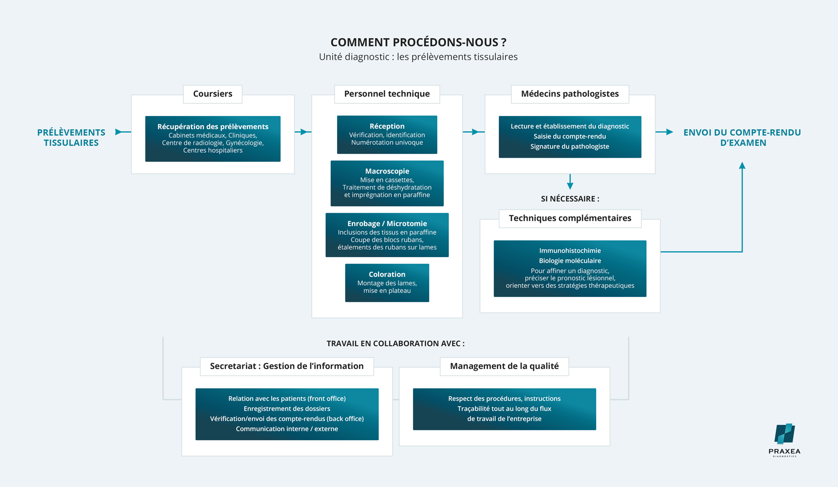
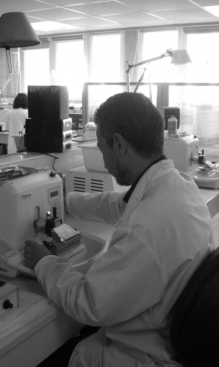
The Histological Examination
The histological examination corresponds to the tissue examination.
The tissue sampling performed on a patient is the result of a clinical decision. Any biopsy or ablation piece has to be analyzed.
The lesion macroscopic description is the first step, followed by a microscopic analysis; this is the standard histopathological examination. If required, the microscopic examination is then performed with more innovative analyses to determine the lesion prognosis (immunohistochemistry, molecular biology techniques).
The use of these techniques complies with the rules of good professional practice and Quality Assurance.
A report is made by the pathologist.
The most common and demonstrative field of application is the study of tumor lesions in cancerology, in order to define these lesions and evaluate their evolutionary potential.
In any case, the histopathological examination is the first step in the care pathway of patients. The histopathological examination is compared with clinical and biological data.
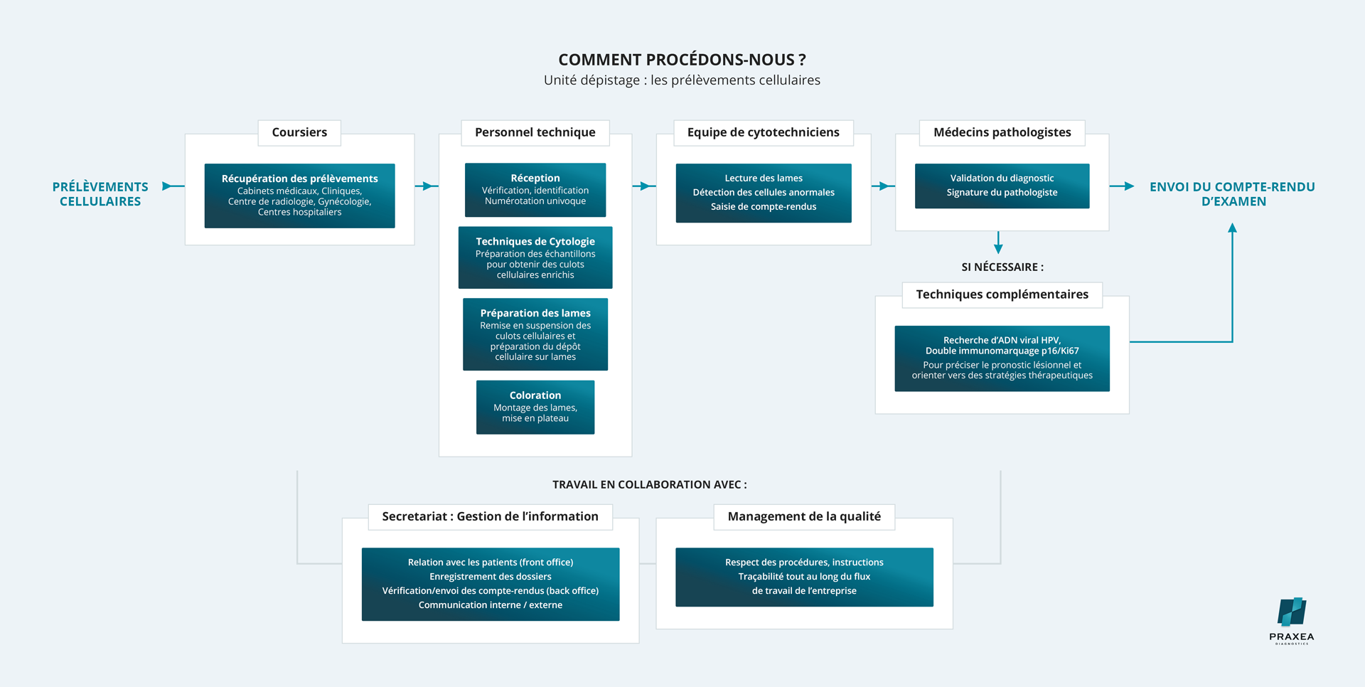
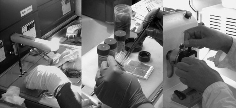
The Cytological Examination
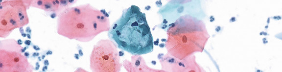
The cytological examination corresponds to the cell examination.
Cells are collected by smear, puncture, suction or straight extraction of biological fluids.
In cervical cancer screening, cells are collected by smear; it is known as the uterine cervical smear.
The cytological sample is processed for the preparation of stained slides, composed of a thin cellular layer. The cells are analyzed under the microscope by pathologists.
Additional techniques can be performed (HPV viral DNA testing, Ki67/p16 dual immunostaining).
Sharing values at the core of our Institute success

Excellence

Innovation

Expertise

Partenariat

Qualité

Index of occupational equality - 99/100
Individual salary increase interval: 35/35
Salary increase of employees returning from maternity leave: 15/15
Parity between the highest salaries: 10/10
About us : Our history

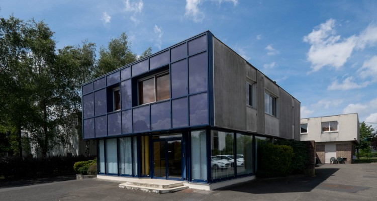
Created in January 2018, Unilabs Praxea Institute of Pathology, located in Massy, near the Plateau de Saclay, has combined the skills of three centers of expertise; the Tolbiac Pathology Office, the Mozart Pathology Office and the "ACP Bièvres - les Ulis" Pathology Center.
The Tolbiac Pathology Office, founded in 2003, is the result of a partnership between four liberal medical structures: the Legland Laboratory, ANA13 (Drs. Ladouch Badre and Triller), Dr. Lefort-Mosse's office and Dr. Gruffaz's office. The team was composed of 7 pathologists and 36 employees.
The “ACP Bièvres - les Ulis” Pathology Center was founded in 1972 in Verrières-le-Buisson by Dr. Baviera and Dr. Guillois. The office was located in Bièvres for 27 years before moving to Les Ulis in 2011. The team was composed of 7 pathologists, with the support of 42 collaborators.
The Mozart Pathology Office was founded in the 1970s by Dr. Demay. Originally located in the 17th arrondissement of Paris, it was located in Clichy, rue Mozart, in 1994, in order to get closer to its partners. The team was composed of 3 pathologists and 15 employees.
Since January 2019, the Unilabs Praxea Institute of Pathology has an entirely new facility in Massy with the latest high-tech equipment, in compliance with Quality Assurance standards. Unilabs Praxea is now composed of a team of 23 pathologists and about a hundred employees.